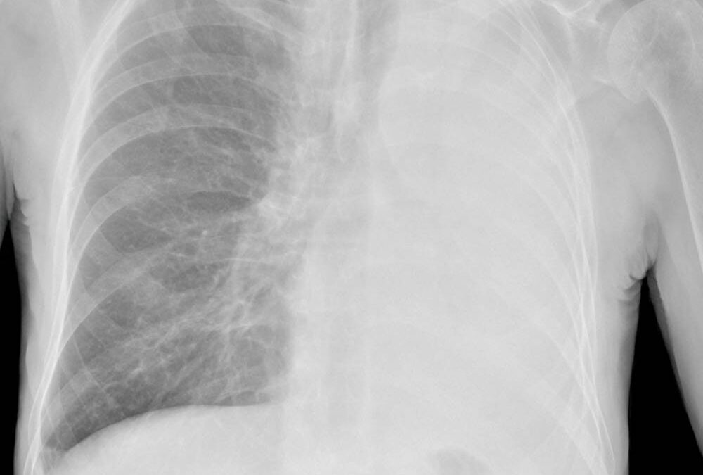
Airway collapse during anesthesia induction is a medical emergency. It can lead to poor ventilation, oxygenation, and in severe cases, death. Both airway trauma and foreign bodies, such as endotracheal tubes, can result in airway collapse by impairing blood flow to the trachea or damaging its structure. Expiratory central airway collapse (ECAC), a degenerative disorder of the trachea and bronchi, is another type of airway emergency that can quickly escalate airway collapse during anesthesia induction (1,2).
Both asthma and COPD are major risk factors and the most common underlying causes of ECAC. The state of chronic inflammation that both diseases cause results in tracheal tissue degeneration and thus lack of structural support for the trachea and bronchi. Typically, collapse of over 70% of the airway has been reported in unexpected airway collapses; however, it is unclear what percentage of airway narrowing predisposes a patient to life-threatening collapse (1). The prodrome of ECAC is often silent until there is severe airway narrowing and collapse. Thus, it is an easy diagnosis to miss and can lead to emergent, life-threatening situations. Initial symptoms can range from cough, dyspnea, and sputum to more severe symptoms like wheezing, coughing or stridor (1). Further, testing for ECAC can be difficult. The only way to truly assess airway collapse is with bronchoscopy or dynamic airway multidetector CT, which is not convenient nor cost effective for a pre-operative workup (4).
When prepping a patient for surgery, it is important for anesthesiologists to be cognizant of any established ECAC diagnosis. Preoperative assessments should be targeted towards assessment of symptoms, as well as prior history of anesthesia complications. Bronchoscopy or CT should be performed and reviewed prior to surgery to assess for the degree of airway collapse in high-risk patients. In cases where collapse is less than 70%, surgery and anesthesia can proceed, but alternatives to general anesthesia are recommended. These include regional anesthesia, neuraxial blocks, or monitored anesthesia care (1). Emergency plans and equipment, such as endotracheal tubes, laryngoscopes, and fiberoptic scopes should be readily available. If airway collapse exceeds 70%, elective surgery should be postponed until corrective ECAC treatment can occur. In emergency surgeries, mechanical circulatory support prior to induction should be considered. This includes mechanical support via machines like cardiopulmonary bypass or ECMO.
In unexpected airway collapse during anesthesia maintenance or induction, intraoperative management should follow a stepwise approach (1). First, physicians should administer 100% oxygen, while checking for obstruction in the endotracheal tube, circuit, or machine. If no obstruction is found, auscultating the lungs for breath sounds is the next step. If no breath sounds are heard and no obstruction is present, a fiberoptic scope can be used to confirm ECAC. Patient positioning can be modified to improve ventilation and slowly advancing the endotracheal tube past the point of collapse to prevent hypoxemia (5). Helium and oxygen can be administered to promote oxygenation and ventilation (6). If hypoxia is unresolved and the patient remains unstable, emergency mechanical circulatory support may be required. Patients who experience ECAC intraoperatively must be carefully monitored postoperatively for respiratory failure after extubation (7).
Overall, ECAC is a difficult diagnosis to make but poses a significant risk for intra-operative emergent airway collapse. Following a stepwise and systematic approach will allow anesthesiologists to adequately manage airway collapse during anesthesia induction and maintenance and ultimately save lives.
References
- Diaz Milian, R., et al. “Expiratory Central Airway Collapse in Adults: Anesthetic Implications (Part 1).” Journal of Cardiothoracic and Vascular Anesthesia, vol. 33, no. 9, Sept. 2019, pp. 2546–2554, 10.1053/j.jvca.2018.08.205.
- Ochs, R. A., et al. “Prevalence of Tracheal Collapse in an Emphysema Cohort as Measured with End-Expiration CT.” Academic Radiology, vol. 16, no. 1, Jan. 2009, pp. 46–53, 10.1016/j.acra.2008.05.020.
- Leong, P., et al. “What’s in a Name? Expiratory Tracheal Narrowing in Adults Explained.” Clinical Radiology, vol. 68, no. 12, Dec. 2013, pp. 1268–1275, 10.1016/j.crad.2013.06.017.
- Ciet, P., et al. “Optimal Imaging Protocol for Measuring Dynamic Expiratory Collapse of the Central Airways.” Clinical Radiology, vol. 71, no. 1, Jan. 2016, pp. e49–e55, 10.1016/j.crad.2015.10.014.
- Kandaswamy, C., and Balasubramanian, V. “Review of Adult Tracheomalacia and Its Relationship with Chronic Obstructive Pulmonary Disease.” Current Opinion in Pulmonary Medicine, vol. 15, no. 2, Mar. 2009, pp. 113–119, 10.1097/mcp.0b013e328321832d.
- Ho, A. M.-H., et al. “Use of Heliox in Critical Upper Airway Obstruction.” Resuscitation, vol. 52, no. 3, Mar. 2002, pp. 297–300, 10.1016/s0300-9572(01)00473-7.
- Popat, M., et al. “Difficult Airway Society Guidelines for the Management of Tracheal Extubation.” Anaesthesia, vol. 67, no. 3, 9 Feb. 2012, pp. 318–340, 10.1111/j.1365-2044.2012.07075.x.


Recent Comments