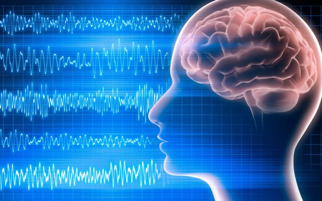
In 1875, electroencephalography was first noted by Dr. Caton, a British physician working at the Liverpool Royal Infirmary. Dr. Caton discovered the presence of electric potentials in the brains of animals using a galvanometer and electrodes (1,2). In 1929, Hans Berger demonstrated that these same electric potentials could also be measured in humans. Last, but not least, in 1937, Gibbs, Gibbs, and Lenox discovered that systematic changes in EEG waveforms could be used to monitor a patient’s response to general anesthesia (2). In this article, the EEG markers for depth of anesthesia associated with propofol, ketamine, and the inhaled anesthetics (sevoflurane, isoflurane, desflurane) will be explored.
Propofol is the most commonly used anesthetic agent and is typically used as an induction agent for sedation and maintenance of general anesthesia (4). Propofol works by binding to postsynaptic GABA receptors, where it induces an inward chloride current which leads to the hyper-polarization and inhibition of postsynaptic neurons (3,4). The EEG patterns observed during propofol-induced anesthesia depend critically on several factors, the most important of which is the rate of drug administration. When propofol is administered as a bolus for induction of anesthesia, the EEG changes within 10-30 seconds from an awake pattern with high-frequency, low-amplitude gamma and beta oscillations to patterns of high-amplitude slow and delta oscillations (4). The appearance of slow and delta EEG oscillations coincides with loss of responsiveness, loss of the oculocephalic reflex, apnea and atonia, serving as markers of reaching sufficient anesthesia depth (4). When the propofol infusion is discontinued, and the patient is allowed to emerge, the slow and alpha oscillations dissipate and are gradually replaced by higher frequency beta and gamma oscillations that have lower amplitudes (4). The return of high-frequency power in the EEG is consistent with return of normal cortical activity and indirectly with return of normal thalamic and brainstem activity. The return of brainstem, thalamic and cortical activity are necessary to restore the awake state (4).
Ketamine is an anesthetic adjunct and analgesic agent that acts primarily as a NMDA receptor antagonist in the brain and the spinal cord. In particular, low-moderate doses of ketamine work to attenuate the inhibitory effects of inhibitory interneurons (3,4). By blocking inputs to inhibitory interneurons, ketamine allows downstream excitatory neurons to become disinhibited or more active. This is why cerebral metabolism increases with low doses of ketamine (4). Additionally, hallucinations and dissociative states are common with low dose ketamine, because brain regions, such as the cortex, hippocampus and the amygdala, continue to communicate but with less modulation and control by the inhibitory interneurons (4). As the dose of ketamine is increased, the NMDA receptors on the excitatory glutamatergic neurons are also blocked and consciousness is lost. The EEG markers associated with ketamine-induced anesthesia are typically fast oscillations in the high beta, low gamma range at between 25 to 32 Hz (4).
The principal inhaled anesthetics are the ether derivatives: sevoflurane, isoflurane, and desflurane. These agents are considered ‘total anesthetics’, as they can maintain all of the required behavioral and physiological characteristics of general anesthesia without adjunct (4). The inhaled agents are known to create their behavioral and physiological effects through binding at multiple targets in the central nervous system, including binding to GABA receptors and enhancing GABAergic inhibition, blockade of 2-pore potassium channels and HCN channels, and blockade of glutamate release via NMDA receptor antagonism (3, 4). At sub-minimal alveolar concentration (MAC) concentrations, sevoflurane creates strong alpha and slow-delta oscillations that closely resemble patterns created by propofol (4). As the concentration of sevoflurane is increased to MAC levels and above, a strong theta oscillation appears in EEG, creating a distinctive pattern of evenly distributed power from the slow oscillation range up through the alpha range and providing a useful marker of reaching the concentration needed for anesthesia (4). Isoflurane and desflurane have similar patterns to sevoflurane. As in the case of sevoflurane, on emergence from isoflurane or desflurane anesthesia, there is loss of the theta oscillation power, followed by dissipation of alpha and slow-delta oscillation power, and reappearance of the power in the beta and gamma bands (4). This relationship between MAC and theta oscillations is useful clinically, as the appearance of theta oscillations indicates a more profound state of unconsciousness and immobility for an inhaled ether anesthetic.
References
- Hajat Z, Ahmad N, Andrzejowski J. The role and limitations of EEG-based depth of anaesthesia monitoring in theatres and intensive care. Anaesthesia. 2017;72 Suppl 1:38-47. doi:10.1111/anae.13739
- Martin JT, FaulconerA Jr, Bickford RG. Electroencephalography in anesthesiology. Anesthesiology. 1959;20(3):359-376. doi:10.1097/00000542-195905000-00017
- Jameson LC, Sloan TB. Using EEG to monitor anesthesia drug effects during surgery. J Clin Monit Comput. 2006;20(6):445-472. doi:10.1007/s10877-006-9044-x
- Purdon PL, Sampson A, Pavone KJ, Brown EN. Clinical Electroencephalography for Anesthesiologists: Part I: Background and Basic Signatures. Anesthesiology. 2015;123(4):937-960. doi:10.1097/ALN.0000000000000841


Recent Comments